It’s pretty common to take photos in our daily lives, but have you ever thought about taking a clear photo of the ions in the portion just inside the cell? Such photo was shot with a resolution under 10 nanometers by the research group led by Academician DU Jiangfeng from University of Science and Technology of China (USTC), cooperating with one led by Academician XU Tao from Chinese Academy of Science (CAS), making it the very first sharp photo of the ferritins, the storage form of iron.
This group independently developed an experimental platform for magnetic imaging (MI) at the nanometer scale for protein in situ (in position), whose central component is the “diamond sensor”, namely, the camera. The sensor is a group of nano pillars made of special diamond that have nitrogen-vacancy center (NV), which are formed by replacing some of the carbon atoms with nitrogen atoms in the diamond structure, resulting in vacancies inside the diamond. In this way the diamond is more sensitive to the faint magnetic signs in the space rendered by the samples.
Then the received signs is processed by the laser and microwave into optical signals which is recognized by the photon detector and imaged by the atomic force microscope (AFM).To get more accurate information, the experimental platform, which carries the cellular sample, is set to get extraordinarily close to the diamond sensor and move in a velocity at the nanometer scale around it, and finally get the image of the molecules inside the cell.
The cell used in this experiment is the hepatic carcinoma cell line (HepG2), which has ferritins, and thus has paramagnetism. To get exact measurement of the active iron ions in the ferritins, the research group used ultrafast, high-pressure freezing to immobilize all intracellular components of the Fe-loaded cells. And then embedded and polymerized these cells inside other compounds with the concerned intracellular structures remaining in situ.
Using a diamond knife, the group then sectioned the tip surface to nanometer flatness to better examine the polymerized cell section, just like making up before taking a photo. The AFM is like the photo booth and the diamond sensor the camera to take photos for the these minor visitors. The research group scanned the cell sample along the diamond nano pillars and acquired MI images of ferritins, whose resolution is on a scale of under 10 nanometers.
This achievement sets an excellent technology basis for further investigation and shows possibility for researches in electron paramagnetic resonance (EPR) spectroscopy at single cell scale.
The MI has been widely used both clinically and laboratorially while the resolution is limited by their electrical detection sensitivity. Thus it’s hard for traditional MI to get sharp photos of biologic molecules in situ at the nanometer scale. The previous study in 2015 of the group has confirmed that MI based on NV in diamonds is capable to detect the EPR spectroscopy of single protein molecule. And in another study in 2018 they realized the probe of the EPR spectroscopy of single DNA molecule in liquid condition.
The first authors of the paper are WANG Pengfei, CHEN Sanyou, GUO Maosen and PENG Shijie from the microscale magnetic resonance central laboratory of USTC. Academician DU Jiangfeng from USTC and Academician XU Tao from CAS are the corresponding authors. And this achievement is published in the Science Advance recently entitled Nanoscale magnetic imaging of ferritins in a single cell.
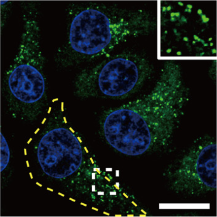
Representative CFM image of ferritin structures (green) in iron-loaded HepG2 cells. The yellow dashed line outlines the contour of a cell. (Image by DU Jiangfeng)
https://advances.sciencemag.org/content/5/4/eaau8038
(Written by LIU Zige, edited by HU Dongyin, USTC News Center)