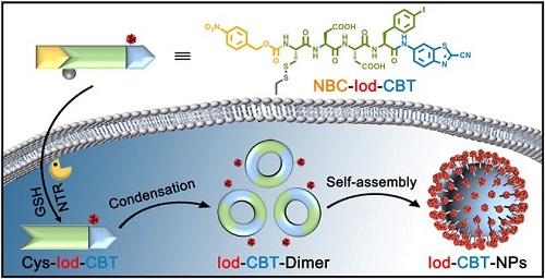
The group led by Prof. LIANG Gaolin from University of Science and Technology of China (USTC) designed an iodine-containing small molecular precursor, which helped in the direct and high contrast observation of the intracellularly formed nanoparticles with Nanocomputed tomography (nano-CT) technique. The result was online published on Science Advances.
Small molecules and nanostructures are showing exciting potentials in molecular imaging and drug delivering. Small molecules are easily taken up by cells but clear fast, while nanostructure has longer retention time in cells but it is more difficult to be taken up by cells.
To combine the advantage of both, researchers developed a smart strategy of incubating cells with a small molecular precursor, which will end up with a nanostructure inside the cell.
Based on their former work of studies on the CBT-Cys click condensation reaction, the team carefully designed NBC-Iod-CBT, an iodinated small-molecule serves as probe.
When taken up by cancer cells, the probe becomes a reactive intermediate after reactions with glutathione and under nitroreductase. Intermediates then condensate through CBT-Cys click reaction, and self-assembly into iodine-rich nanoparticles.

Schematic illustration on how probe NBC-Iod-CBT self-assembly into the iodine-rich nanoparticles (Iod-CBT-NPs) inside a cell. (Image by ZHANG Miaomiao)
However, as some other nanostructures are intrinsic inside the cell, it remains challenging to distinguish between the two.
Traditional observation techniques are not ideal for observing the 3D nanostructures in the whole cell either due to solution limitation or poor performance in imaging thick sample.
Nano-CT meets the demands. Water window technology of nano-CT allows an unstained, around 10-um-thick, frozen-hydrated cell to be 3D imaged in its near native state with unique high contrast and resolution.
By nano-CT, researchers were able to directly observe spatial distribution of the cellular organelles as well as the artificially formed iodine-rich nanostructures with high contrast while the cryopreserved cells not being sliced.
Moreover, because different substances have different X-ray absorption capabilities and their linear absorption coefficient values are different, the nanoparticles formed in the cell can be further determined by their linear absorption coefficient.
This strategy developed is expected to help people further distinguish other artificial nanostructures formed in cells, so as to gain insight into the formation mechanism of nanostructures in cells.
(Written by JIANG Pengcen, edited by HU Dongyin, USTC News Center)